Cataract surgery is a procedure to remove the lens of your eye and, in most cases, replace it with an artificial lens. Normally, the lens of your eye is clear. A cataract causes the lens to become cloudy, which eventually affects your vision.
Cataract surgery is performed by an eye doctor (ophthalmologist) on an outpatient basis, which means you don’t have to stay in the hospital after the surgery. Cataract surgery is very common and is generally a safe procedure.
Why it’s done
Cataract surgery is performed to treat cataracts. Cataracts can cause blurry vision and increase the glare from lights. If a cataract makes it difficult for you to carry out your normal activities, your doctor may suggest cataract surgery.
When a cataract interferes with the treatment of another eye problem, cataract surgery may be recommended. For example, doctors may recommend cataract surgery if a cataract makes it difficult for your eye doctor to examine the back of your eye to monitor or treat other eye problems, such as age-related macular degeneration or diabetic retinopathy.
In most cases, waiting to have cataract surgery won’t harm your eye, so you have time to consider your options. If your vision is still quite good, you may not need cataract surgery for many years, if ever.
When considering cataract surgery, keep these questions in mind:
- Can you see to safely do your job and to drive?
- Do you have problems reading or watching television?
- Is it difficult to cook, shop, do yardwork, climb stairs or take medications?
- Do vision problems affect your level of independence?
- Do bright lights make it more difficult to see?
Risks
Complications after cataract surgery are uncommon, and most can be treated successfully.
Cataract surgery risks include:
- Inflammation
- Infection
- Bleeding
- Swelling
- Drooping eyelid
- Dislocation of artificial lens
- Retinal detachment
- Glaucoma
- Secondary cataract
- Loss of vision
Your risk of complications is greater if you have another eye disease or a serious medical condition. Occasionally, cataract surgery fails to improve vision because of underlying eye damage from other conditions, such as glaucoma or macular degeneration. If possible, it may be beneficial to evaluate and treat other eye problems before making the decision to have cataract surgery.
How you prepare
Food and medications
You may be instructed not to eat or drink anything 12 hours before cataract surgery. Your doctor may also advise you to temporarily stop taking any medication that could increase your risk of bleeding during the procedure. Let your doctor know if you take any medications for prostate problems, as some of these drugs can interfere with cataract surgery.
Antibiotic eyedrops may be prescribed for use one or two days before the surgery.
Other precautions
Normally you can go home on the same day as your surgery, but you won’t be able to drive, so arrange for a ride home. Also arrange for help around home, if necessary, because your doctor may limit activities, such as bending and lifting, for about a week after your surgery.
What you can expect
Before the procedure
A week or so before your surgery, your doctor performs a painless ultrasound test to measure the size and shape of your eye. This helps determine the right type of lens implant (intraocular lens, or IOL).
Nearly everyone who has cataract surgery will be given IOLs. These lenses improve your vision by focusing light on the back of your eye. You won’t be able to see or feel the lens. It requires no care and becomes a permanent part of your eye.
A variety of IOLs with different features are available. Before surgery, you and your eye doctor will discuss which type of IOL might work best for you and your lifestyle. Cost may also be a factor, as insurance companies may not pay for all types of lenses.
IOLs are made of plastic, acrylic or silicone. Some IOLs block ultraviolet light. Some IOLs are rigid plastic and implanted through an incision that requires several stitches (sutures) to close.
However, many IOLs are flexible, allowing a smaller incision that requires few or no stitches. The surgeon folds this type of lens and inserts it into the empty capsule where the natural lens used to be. Once inside the eye, the folded IOL unfolds, filling the empty capsule.
Some of the types of lenses available include:
- Fixed-focus monofocal. This type of lens has a single focus strength for distance vision. Reading will generally require the use of reading glasses.
- Accommodating-focus monofocal. Although these lenses only have a single focusing strength, they can respond to eye muscle movements and shift focus to near or distant objects.
- Multifocal. These lenses are similar to glasses with bifocal or progressive lenses. Different areas of the lens have different focusing strengths, allowing for near, medium and far vision.
- Astigmatism correction (toric). If you have a significant astigmatism, a toric lens can help correct your vision.
Discuss the benefits and risks of the different types of IOLs with your eye surgeon to determine what’s best for you.
During the procedure
Cataract surgery, usually an outpatient procedure, takes an hour or less to perform.
First, your doctor will place eyedrops in your eye to dilate your pupil. You’ll receive local anesthetics to numb the area, and you may be given a sedative to help you relax. If you’re given a sedative, you may remain awake, but groggy, during surgery.
During cataract surgery, the clouded lens is removed, and a clear artificial lens is usually implanted. In some cases, however, a cataract may be removed without implanting an artificial lens.

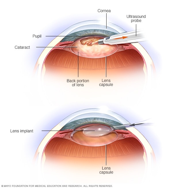
During phacoemulsification — the most common type of cataract surgery — the rapidly vibrating tip of the ultrasound probe emulsifies and helps break up the cataract, which your surgeon then suctions out (top). An outer housing of the cataract (the lens capsule) is generally left in place. After removing the emulsified material, your surgeon inserts the lens implant into the empty space within the capsule where the natural lens used to be (bottom).[/ultimate_modal]
Click image for more info.
Surgical methods used to remove cataracts include:
- Using an ultrasound probe to break up the lens for removal. During a procedure called phacoemulsification (fak-o-e-mul-sih-fih-KAY-shun), your surgeon makes a tiny incision in the front of your eye (cornea) and inserts a needle-thin probe into the lens substance where the cataract has formed.Your surgeon then uses the probe, which transmits ultrasound waves, to break up (emulsify) the cataract and suction out the fragments. The very back of your lens (the lens capsule) is left intact to serve as a place for the artificial lens to rest. Stitches may be used to close the tiny incision in your cornea at the completion of the procedure.
- Making an incision in the eye and removing the lens in one piece. A less frequently used procedure called extracapsular cataract extraction requires a larger incision than that used for phacoemulsification. Through this larger incision your surgeon uses surgical tools to remove the front capsule of the lens and the cloudy lens comprising the cataract. The very back capsule of your lens is left in place to serve as a place for the artificial lens to rest.This procedure may be performed if you have certain eye complications. With the larger incision, stitches are required.
Once the cataract has been removed by either phacoemulsification or extracapsular extraction, the artificial lens is implanted into the empty lens capsule.
After the procedure
After cataract surgery, expect your vision to begin improving within a few days. Your vision may be blurry at first as your eye heals and adjusts.
Colors may seem brighter after your surgery because you are looking through a new, clear lens. A cataract is usually yellow- or brown-tinted before surgery, muting the look of colors.
You’ll usually see your eye doctor a day or two after your surgery, the following week, and then again after about a month to monitor healing.
It’s normal to feel itching and mild discomfort for a couple of days after surgery. Avoid rubbing or pushing on your eye.
Your doctor may ask you to wear an eye patch or protective shield the day of surgery. Your doctor may also recommend wearing the eye patch for a few days after your surgery and the protective shield when you sleep during the recovery period.
Your doctor may prescribe eyedrops or other medication to prevent infection, reduce inflammation and control eye pressure. Sometimes, these medications can be injected into the eye at the time of surgery.
After a couple of days, most of the discomfort should disappear. Often, complete healing occurs within eight weeks.
Contact your doctor immediately if you experience any of the following:
- Vision loss
- Pain that persists despite the use of over-the-counter pain medications
- Increased eye redness
- Eyelid swelling
- Light flashes or multiple new spots (floaters) in front of your eye
Most people need glasses, at least some of the time, after cataract surgery. Your doctor will let you know when your eyes have healed enough for you to get a final prescription for eyeglasses. This is usually between one and three months after surgery.
If you have cataracts in both eyes, your doctor usually schedules the second surgery after the first eye has healed.
Results
Cataract surgery successfully restores vision in the majority of people who have the procedure.
People who’ve had cataract surgery may develop a secondary cataract. The medical term for this common complication is known as posterior capsule opacification (PCO). This happens when the back of the lens capsule — the part of the lens that wasn’t removed during surgery and that now supports the lens implant — becomes cloudy and impairs your vision.
PCO is treated with a painless, five-minute outpatient procedure called yttrium-aluminum-garnet (YAG) laser capsulotomy. In YAG laser capsulotomy, a laser beam is used to make a small opening in the clouded capsule to provide a clear path through which the light can pass.
After the procedure, you usually stay in the doctor’s office for about an hour to make sure your eye pressure doesn’t rise. Other complications are rare but can include increased eye pressure and retinal detachment.

